
PET/CT (Positron-Emission-Tomography/Computed-Tomography)
MRI (Magnetic-Resonance-Imaging)
PET/CT imaging is performed by injection of a very small amount of radioactive tracer (e.g. PSMA or FDG depending on the kind of disease/tumor). After an uptake time of 60 minutes the PET/CT-scan will be performed. PET/CT enables detection of very small tumor deposits (within the millimeter-range) and localize them exactly in the body. PET/CT-imaging is increasingly used in modern cancer therapy to define an optimal individual treatment concept for the patient.
MRI is made without radiation. MRI scanners use magnetic fields and radio waves to form images of the body. Unlike x-ray examinations and computed tomography (CT) scans, MRI does not utilize on ionizing radiation. Instead, radio waves redirect alignment of hydrogen atoms that exist within the body while you are in the scanner without causing any chemical changes in the tissues. As the hydrogen atoms return to their usual alignment, they emit energy that differs according to the type of tissue from which they come. The MR scanner captures this energy and creates precise and high-resolution cross-sectional images of the tissues scanned. Many different body parts and organs can be examined in detail. That is why this imaging technology has spread and established worldwide. A Modern MRI unit is a large cylinder-shaped tube surrounded by a circular magnet, in the middle of which the patient must be positioned to obtain optimal image quality. We also offer MRI machines with a larger diameter bore (70 cm) which can be more comfortable for larger size patients or patients with claustrophobia. Furthermore, the presence of an accompanying person in the examination room or the application of a calmative agent may be helpful to perform the examination. Please inform us when making an appointment if you suffer from claustrophobia / agoraphobia.
Please bring the following to the examination:
- referral and chip card
- Current creatinine level, if you have a kidney disease, diabetes mellitus or if you are older than 50 years
- Previous images
- Patients with medical implants (except joint prostheses) are asked to bring their equipment license, which certify the MRI compatibility if applicable
- X-ray and allergy passport (if available)
Functional prostate MRI
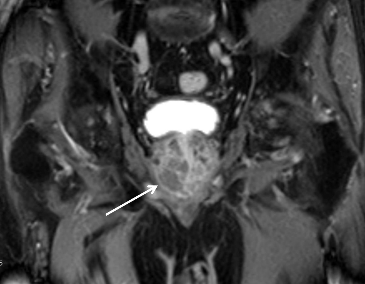
The functional prostate MRI uses special test parameters (Dynamic Contrast-aided sequences, Diffusion-weighted sequences, high-resolution T2-weighted sequences) that can accurately represent the location and extent of prostate cancer. This is important for treatment planning. This method is used in elevated PSA levels when biopsies could not prove a tumor, but there is reason for the suspicion of prostate cancer. Even after surgery and radiotherapy of prostate cancer, the functional prostate MRI can detect a local recurrence of the tumor or exclude it with high probability.
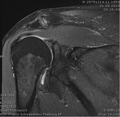
Upper arm / shoulder joint in MRI
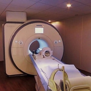
MR Scanner
T2 - weighted MRI of the prostate
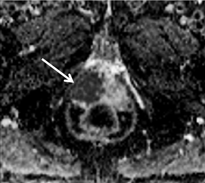
Diffusion - weighted MRI: the dark spot (arrow ) marks the prostate cancer

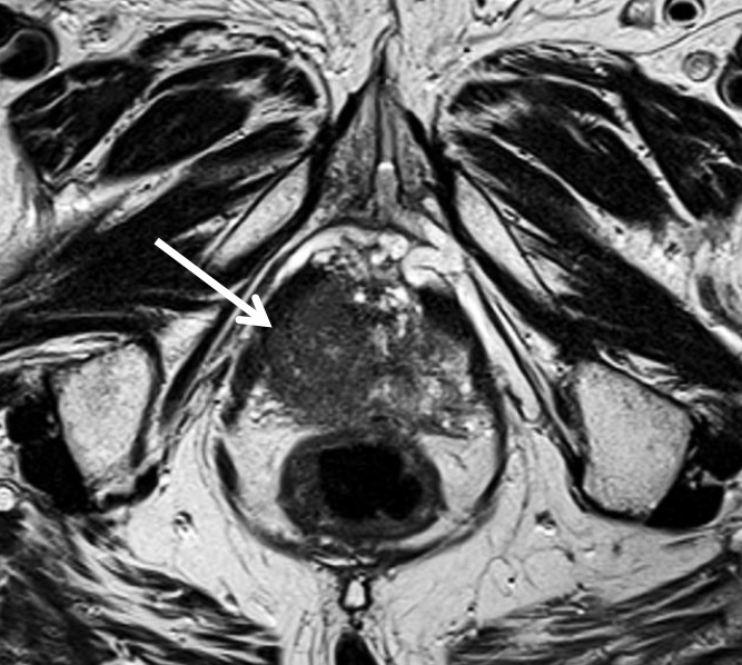
T2 - weighted MRI of the prostate: the arrow marks the cancer
MRI of the prostate and pelvis after dynamic application of contrast medium: the tumor in the prostate (arrow) shows a rapid washout of contrast medium. No evidence of lymph node metastases